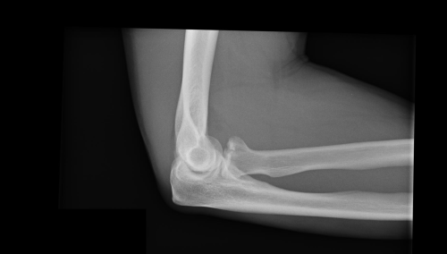
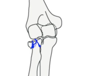
Radiuskop fractuur
Radiuskop fractuur
Auteur: J. Sprakel, MD - Laatste update: 18-06-2014
Radiuskop fractuur
Lichamelijk onderzoek
- Kliniek
- - Pijn aan laterale zijde van elleboog
- - Functio laesa elleboog
- - Geringe zwelling van elleboog
- - Drukpijn over radiuskop
- - Pijn bij pro- en supinatie
- - Pijn neemt toe bij pronatie
- - Pols altijd onderzoeken ! (cave: scheur membrana interossea - Essex Lopresti)
- Begeleidende letsels:
- - Essex Loprestie letsel (radiuskop/halsfractuur met distale radio-ulnaire luxatie)
- - Elleboogluxatie
- - Olecranonfractuur
- - Ruptuur van ulnaire collaterale ligamenten
Classificatie
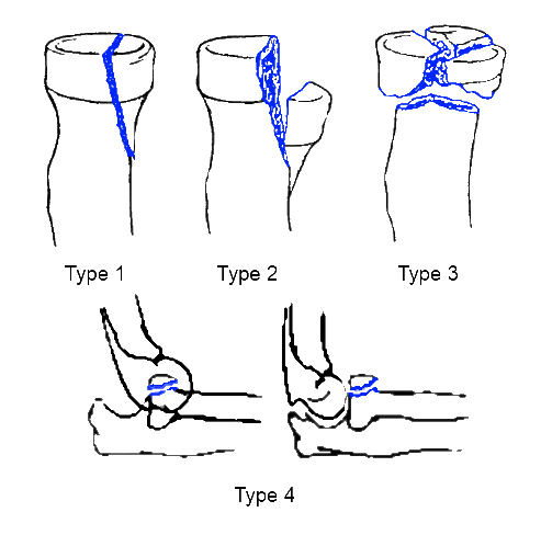
Classificatie volgens Broberg-Morrey Modification of the Mason Classification 1
| Type | Beschrijving |
|---|---|
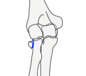
| Type 1 | Rand van radiuskop, <2mm gedisloceerde fractuur |
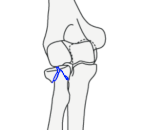
| Type 2 | Beitelfractuur("Meissel Fraktur"), >2mm gedisloceerde fractuur, niet comminutief, reconstrueerbaar |
| Type 3 | Comminutieve fractuur, niet reconstrueerbaar |
| Type 4 | In combinatie met elleboogluxatie |
Classificatie volgens Broberg-Morrey Modification of the Mason Classification
| Classificatie volgens AO | ||
| Één bot intra-articulair | ||
|---|---|---|
| 21-B2 Geïsoleerde radius | ||
AO 21-B2.1 Radiuskopfractuur |
AO 21-B2.2 Multifragmentaire radiuskopfractuur zonder gewrichtsdepressie |
AO 21-B2.3 Multifragmentaire radiuskopfractuur met gewrichtsdepressie |
Conservatieve behandeling
Indicaties:- - Mason Type 1
(Na-)behandeling:
- - Eventuele gewrichtspunctie in verband met haemarthros
- - Drukverband gedurende 5-7 dagen, bij veel pijn bovenarmgips voor 1 week
- - Actief oefenen na 1 week
Follow-up:
- - Poliklinische controle na 1 week met oefeninstructies
- - Poliklinische controle na 4 weken met functiecontrole
Functiecontrole:
 |
 |
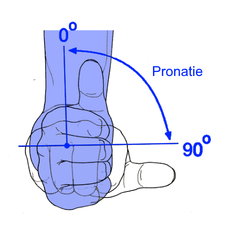 |
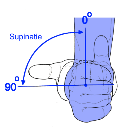
| Flexie / Extensie | Supinatie bij elleboog in 90° flexie | Pronatie bij elleboog in 90° flexie |
| 150° - 0° - 10° | 90° – 0° – 90° | |
Operatieve behandeling
Indicaties:- - Mason Type 2
- - Mason Type 3
(Na-)behandeling:
- - Postero-laterale benadering (Kocher) tussen m. extensor carpi ulnaris en m. anconeus
- - Mason Type 2: Schroeffixatie AO mini fragmentschroef
- - Mason Type 3: Primaire/secundaire (na 3-6 weken) radiuskopextirpatie met prothese
- - Bij zeer comminutieve fracturen bij jonge patienten (<50 jaar) zeer terughoudend met radiuskopextirpatie
- - Brace bij instabiliteit voor 6 weken
Follow-up:
- - Poliklinische controle na 1 weken week met X-elleboog AP en lateraal met oefeninstructies
- - Poliklinische controle na 3 weken met X-elleboog AP en lateraal
- - Poliklinische controle na 6 weken met X-elleboog AP en lateraal met functiecontrole
- - Prothese levenslang controleren
Functiecontrole:
|
|
|
| Flexie / Extensie | Supinatie bij elleboog in 90° flexie | Pronatie bij elleboog in 90° flexie |
| 150° - 0° - 10° | 90° – 0° – 90° | |


Coderingen
Diagnose Behandel Combinatie (DBC/DOT)
ICD-10
| Chirurgie | 210 |
| Orthopedie | 3011 |
ICD-10
| Fractuur van proximale radius | S52.1 |
Abbreviated Injury Scale (AIS)
| Arm fracture NFS | 751800.2 |
| Arm fracture - open | 751801.2 |
| Forearm fracture NFS | 751900.2 |
| Forearm fracture - open | 751901.2 |
| Proximal radius fracture | 752111.2 |
| Proximal radius fracture - open | 752112.2 |
| Proximal radius fracture - partial articular: radial head | 752161.2 |
| Proximal radius fracture - partial articular: radial head - open | 752162.3 |
| Proximal radius fracture - complete articular | 752171.2 |
| Proximal radius fracture - complete articular - open | 752172.3 |