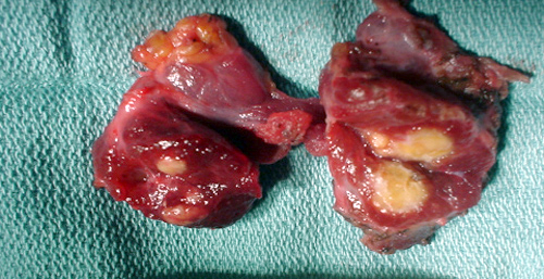
Schildkliercarcinoom
Schildkliercarcinoom
Auteur: J. Sprakel, MD - Laatste update: 25-04-2018
Schildkliercarcinoom

Algemeen
- Het betreft hier de richtlijn over het goed gedifferentieerd schildkliercarcinoom.
- Goed gedifferentieerd schildkliercarcinoom (folliculair epitheel) (80-85%):
- - Papillair carcinoom
- - Folliculair carcinoom / Hürthle cel carcinoom
- - Verhouding papillair : folliculair van 4:1
- - Incidentie: man 2/100.000 per jaar - vrouw 4,5/100.000 per jaar
- Ongedifferentieerd schildkliercarcinoom (15-20%):
- - Medullair carcinoom - C-cellen (5-10%)
- - Anaplastisch/ongedifferentieerd carcinoom (6%)
Kliniek
- Kenmerken die meer voorkomen bij maligniteit:
- - Nieuwe nodus of een die duidelijk toeneemt in grootte
- - Nodus bij een positieve familie-anamnese voor schildkliercarcinoom
- - Nodus bij patienten met bestraling van de hals in de voorgeschiedenis met name op jeugdige leeftijd
- - Nodus bij patienten <20 of >60 jaar en speciaal bij mannen
- - Onverklaarde heesheid en verandering van stem geassocieerd met een struma
- - Cervicale lymfadenopathie (met name diep cervicaal of supraclaviculair)
- - Stridor (laat symptoom)
- Lichamelijk onderzoek:
- - Afwijking meestal >1,5cm
- - Enkelvoudig of multinodulair struma
- - Fixatie van solide nodus aan de omgeving
- - Pathologische lymfeklieren
Diagnostiek
- Laboratorium onderzoek
- - TSH-bepaling (afwijkend --> verwijzing internist-endocrinoloog)
- Echo hals + US-FNAC
- - Echogeleide Fine Needle Aspiration Cytology (US-FNAC) (in 8-20% van de gevallen onvoldoende materiaal) 1-4
- - Bij onvoldoende materiaal herhaal echogeleide FNAC (in 3-4% van de gevallen onvoldoende materiaal) 2,3,5
- Immuuncytochemie
- - Over het algemeen geen indicatie
- - Bij DD schildkliercarcinoom of lymfoom (LCA, thyreoglobuline en TTF1)
- - Aantonen/uitsluiten medullair schildkliercarcinoom (clacitonine, CEA, thyreoiglobuline)
- Histologisch naaldbiopt:
- - Over algemeen geen aanvullende waarde boven cytologie
- - Alleen bij sterke verdenking op anaplastisch carcinoom
- Incidentaloom schildklier (niet palpabele nodus)
-
- Bij echografie: Geen routinematige diagnostiek - Bij CT of MRI: Geen routinematige diagnostiek - Bij FDG-PET(/CT): FNAC bij verhoogd TSH - Redenen om wel diagnostiek te verrichten: 1. Ongerustheid bij patient 2. Combinatie van beeldvormende parameters die voor de onderzoeker reden zijn om voor nadere diagnostiek te kiezen
Bethesda classificatie
Bethesda classificatie voor schildklier cytologie 6-7| Bethesda | Categorie | Beschrijving | Kans op maligniteit | Management suggesties |
|---|---|---|---|---|
| 1 | Niet-diagnostisch |
- Cyste-inhoud - Acellulair preparaat - Overig (klontering, bloed; tevens < 6 groepen van 10 duidelijke folliculaire cellen, gedegenereerde, slecht kleurende folliculaire cellen) |
1-4% | Herhalen FNAC met echografie |
| 2 | Benigne |
- Benigne folliculaire nodus (adenomatoid, hyperplastisch, colloid, etc) - Lymfocytaire (Hashimoto) thryreoiditis - Granulomateuze (subacute) thryreoiditis |
0-3% | Klinische follow-up |
| 3 | AUS-FLUS | - Atypie of folliculaire laesie van onzekere betekenis | 5-15% | Herhalen FNAC met echografie |
| 4 | Folliculaire neoplasie |
- Verdacht voor folliculaire neoplasie - Specificeer bij Hürthle cel (oncocytair) type |
15-30% | Diagnostische hemithyreoidectomie |
| 5 | Verdacht voor maligniteit | Verdacht voor: - papillair schildkliercarcinoom - medullair schildkliercarcinoom - metastase - maligne lymfoom |
60-75% | Diagnostische hemi- / Totale thyreoidectomie |
| 6 | Maligne |
- Papillair schildkliercarcinoom - Slecht gedifferentieerd schildkliercarcinoom - Medullair schildkliercarcinoom - Ongedifferentieerd (anaplastisch) schildkliercarcinoom - Plaveiselcelcarcinoom - Metastase - Non-Hodgkin lymfoom |
97-99% | Totale thyreoidectomie |
TIRADS
TI-RADS (Thyroid Imaging Reporting and Data System-classification) classificatie wordt gebruikt in radiologische verslaglegging 8-9| TI-RADS | Beschrijving | Advies | Kans op maligniteit |
|---|---|---|---|
| 0 | Onvolledig onderzoek | Nieuwe beeldvorming of vergelijking met voorgaand onderzoek noodzakelijk. | |
| 1 | Normaal schildklierweefsel | Geen afwijkingen aantoonbaar | |
| 2 | Benigne laesie | Er wordt een benigne afwijking gezien | |
| 3 | Waarschijnlijk benigne laesie | Waarschijnlijk benigne laesie. Aanvullende punctie of een controle na 6 maanden. | <5% |
| 4A | Mild verdacht | Waarschijnlijk maligne laesie. Aanvullende punctie moet verricht worden. | 5% - 10% |
| 4B | Matig verdacht | Waarschijnlijk maligne laesie. Aanvullende punctie moet verricht worden. | 10% - 80% |
| 4C | Ernstig verdacht | Waarschijnlijk maligne laesie. Aanvullende punctie moet verricht worden. | 10% - 80% |
| 5 | Waarschijnlijk maligne laesie | Zeer verdacht voor maligniteit. Aanvullende punctie moet verricht worden. | >80% |
| 6 | Biopsie bewezen maligniteit | Bijv. bij beeldvorming ter beoordeling effect neoadjuvante therapie. | 100% |
TNM-classificatie & pTNM classificatie
TNM classificatie voor papillair en folliculair schildkliercarcinoom| T - primaire tumor | N - Regionale lymfeklieren | M - Afstandsmetastasen | |||
|---|---|---|---|---|---|
| Tx | Primaire tumor kan niet worden geidentificeerd | Nx | Regionale lymfeklieren kunnen niet worden geidentificeerd | ||
| T0 | Geen aanwijzingen voor een primaire tumor | N0 | Geen aanwijzingen voor regionale lymfekliermetastasen | M0 | Geen afstandsmetastasen |
| T1a | Tumor <1cm, gelimiteerd tot de schildklier | N1a | Lymfekliermetastase in level VI (pre- en paratracheaal, inclusief prelaryngeaal en Delphian lymfeklieren | M1 | Afstandsmetastasen |
| T1b | Tumor >1cm en <2cm, gelimiteerd tot de schildklier | N1b | Lymfekliermetastasen in andere unilaterale, bilaterale of contralaterale cervicale of bovenste medistainale lymfeklieren | ||
| T2 | Tumor >2cm en <4cm, gelimiteerd tot de schildklier | ||||
| T3 | Tumor >4cm, gelimiteerd tot de schildklier of met minimale uitbreiding extra-thyroidaal (e.g. uitbreiding in sternothyroidale spier of perithyroidale weke delen) | ||||
| T4a | Tumor groeit buiten schildklier en groeit in één van de volgende structuren: subcutane weke delen, larynx, trachea, oesophagus, n. laryngeus recurrens (tak van n. vagus) | ||||
| T4b | Tumor groeit in pre-vertebrale fascie, mediastinale vaten of omhult de carotis | ||||
| Alle anaplastische carcinomen zijn T4 tumoren | |||||
| T4a | Tumor van elke grootte, gelimiteerd tot de schildklier | ||||
| T4b | Tumor van elke grootte, met groei buiten de schildklier | ||||
Behandeling
- (Diagnostische) Hemithyreodectomie:
- - Bethesda 4 & 5
- - Papillair schildkliercarcinoom <1 cm zonder aanwijzingen op lymfekliermetastasen
- Totale thyreodectomie gevolgd door 131I:
- - Bethesda 5 & 6
- - Niet radicale resectie na hemithreodectomie
- - Multifocaal papillair carcinoom in het hemithyreoidectomie preparaat
- - Patienten met verhoogde kans op schildkliermaligniteit in contralaterale schildklierkwab
- - Minimaal invasief folliculair carcinoom (MIFC)
Referenties
- 1. Alexander EK, Heering JP, Benson CB, Frates MC, Doubilet PM, Cibas ES, et al.Assessment of nondiagnostic ultrasound-guided fine needle aspirations of thyroid nodules. J Clin Endocrinol Metab 2002; 87(11):4924-7.
- 2. Al Maqbali T, tedla M, Weickert MO, Mehanna H. Malignancy risk analysis in patiënts with inadquate fine needle aspiration cytology (FNAC) of the thyroid. PloS ONE 2012; 7(11): e49078.
- 3. Chow LS, Gharib H, Goellner JR, van Heerden JA.Nondiagnostic thyroid fine-needle aspiration cytology: management dilemmas.Thyroid 2001; 11(12)1147-1151.
- 4. Choi YS, Hong SW, Kwak JY, Moon HJ, Kim EK Clinical and ultrasonographic findings affecting nondiagnostic results upon the second needle aspiration for thyroid nodules. Ann Surg Oncol 2012; 19: 2304-2309.
- 5. Danese D, Sciacchitano S, Farsetti A, Andreoli M, Pontecorvi A. Diagnostic accuracy of conventional versus sonography-guided fine-needle aspiration biopsy of thyroid nodules. Thyroid 1998; 8(1):15-21.
- 6. Rossi ED, Raffaelli M, Mule A, Zannoni GF, Pontecorvi A, Santeusanio G, Minimo C, Fadda G. Relevance of immunohistochemiostry on thin-layer cytology in thyroid lesions suspicious for medullary carcinoma. A case-control study. Appl Immunohistochem Mol Morphol 2008; 16: 548-553.
- 7. Cibas ES, Ali SZ. The 2017 Bethesda System for Reporting Thyroid Cytopathology. Thyroid. 2017 Nov;27(11):1341-1346.
- 8. Horvath E, Majlis S, Rossi R et-al.An ultrasonogram reporting system for thyroid nodules stratifying cancer risk for clinical management. J. Clin. Endocrinol. Metab. 2009;94 (5): 1748-51.
- 9. Kwak JY, Han KH, Yoon JH et-al.Thyroid imaging reporting and data system for US features of nodules: a step in establishing better stratification of cancer risk. Radiology. 2011;260 (3): 892-9.
- 1.